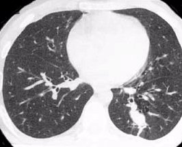
a CT scan with left lower lobe lesion (not mine)
Dr. M, emailed me the preliminary report from the radiologist yesterday afternoon with the comment "good news for your weekend (preliminary)". I've highlighted what I think are the best parts of the report. Here it is:
Findings:
Lungs: An ill defined spiculated lesion is seen in the posterior basal segment of left lower lobe abutting the descending thoracic aorta. It measures 2 x 2 cm on image 3/87, previously measured 2.7 x 1.8 cm on that CT dated October 6, 2009 and 2.8 x 1.8 cm on CT dated
September 16, 2009. 2nd pleural-based ill-defined lesion in the posterior basal segment of left lower lobe measures 10 x 9 mm on image 3/59, previously measured 17 x 11 mm (October 6, 2009) 14 x 12 mm (CT study dated September 16, 2009).
Multiple calcified nodules are present in both lungs. No new pulmonary nodule.
Mediastinum: Multiple small mediastinal lymph nodes, unchanged. Tip of the Port-A-Cath is seen at the cavoatrial junction.
Heart size is normal.
No pericardial or pleural effusions.
Trachea and bronchi are patent.
Included portions of the abdominal reveal diverticuli at the hepatic flexure. Multiple calculi are also present in the gallbladder.
No lytic and blastic osseous lesions.
Impression:
1. Interval decrease in the size of left lower lobe lesions.
2. No new lesion concerning for disease progression.
3. Cholelithiasis.
No lytic and blastic osseous lesions means it hasn't spread to my bones. So to begin with there is much happiness and rejoicing around these parts.
However, I continue to be perplexed about how to really understand the CT scans from my non-medical viewpoint. This may be undue worries on my part, but they exist nonetheless. Let me try to explain.
I've had 7 CT scans and 1 PET scan in the past year. They are read by radiologists, who are highly skilled and trained to do this. I look at the scans and see vague shapes and black and white spots. Radiologists can make sense of the scans--they can pick out what spots are likely cancerous or not. Another part of radiologists' training includes learning a highly specialized vocabulary that is not in common usage among non-physicians. The CT scans are read by the doctor who is on duty that day, so the readings may be influenced by radiologist who is doing the reading.
In point of fact, each of my CT scans reads a little bit differently, with respect to who the radiologist is and what the radiologist has selected to view. The "ill defined spiculated lesion" at the bottom and back of my left lower lung lobe, identified by yesterday's read, was not mentioned whatsoever in my CT scans of 7/15/10; 4/28/10; 2/19/10; and 12/9/09. I'm wondering where this tumor went during those times because it is the largest of all the identified tumors in my lungs. And the 10/6/09 scan was a PET scan not a CT scan, just to clear the record.
I plan to ask Dr. M about this discrepancy when I see him on Tues prior to my next chemotherapy infusion. I know that my training as a lawyer, where language is honed to a precise level and consistency is drummed into attorneys in training as a virtue, can clash with the complex and sometimes messy nature of human existence and observation. And, as this CT scan was the first of mine done at SCCA, the radiologist may not have had all the records from Group Health available for the comparison. All the same I would like to know why the spiky abnormal tissue seen at the bottom rear of my left lower lung seems to have disappeared from view for 4 of my CT scans.


3 comments:
It's all gobbledegook to me - as an English teacher and a writer, that freaks me a little. But hurray for the shrinking.
Thinking of you.
Zebaxx
Yay, moe!
You realize, don't you, that Twiggy was wearing false eyelashes? That you may buy yourself today at any drug store? Now is the time!!
And the cartoon is great.
Love,
Mel
Post a Comment